3 Brightfield Microscopy
Primary Parts and Light Path
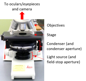
In an upright, transmitted-light microscope (Fig. 1), light is generated by the light source. The condenser lens collects the light and focuses it on the specimen on the stage. The objective lens collects light that makes it through the specimen and sends the light to the eyepieces (oculars) and/or the camera. If you are using an inverted microscope, the light path is exactly the same, but everything is upside down so that the light source and condenser are on the top of the microscope and the objectives are underneath the stage, pointing up. Proper alignment of the microscope optics (a state called Koehler Illumination) is essential for obtaining a good image. You will learn the alignment procedure during your training.
The Basis for Contrast in Brightfield Microscopy
Specimen objects are visible in brightfield microscopy because they absorb the light, resulting in a localized decrease in light intensity. An object that partially attenuates the light appears gray; an object that does not allow any light to pass appears black. Light that travels through empty areas of the slide/dish or through objects that do not absorb light appears bright. Therefore, in brightfield microscopy, objects appear dark against a light background.
Colored objects appear colored because they absorb different wavelengths of light in different amounts. The light produced by the light source contains a broad spectrum of colors (wavelengths) of light (Fig. 2). An object that absorbs blue and green light and allows other colors to pass will appear orange-red.

Many thin/small biological specimens such as cells and tissue sections do not absorb nearly any light (i.e. they are essentially “clear”) and are therefore very difficult to see using brightfield microscopy. Such specimens are often stained with colored dyes (e.g. H&E, Gram stain), making them absorb more light so that they can be seen. Most commonly used stains also aid in the identification of specific cell or tissue features.
Alternatively, a specimen can be stained with fluorescent dyes and then be visualized using fluorescence microscopy. Unstained specimens can be visualized using a more advanced transmitted-light technique such as phase contrast microscopy or differential interference microscopy (DIC).
