5 Differential Interference Contrast (DIC) Microscopy
Differential Interference Contrast (DIC) microscopy is a technique to visualize specimens that do not absorb light and are therefore not visible using standard brightfield microscopy. Although many biological specimens do not absorb light, they do temporarily slow it down. The speed of light in a given medium is slower than the speed of light in a vacuum and this difference is quantified by the medium’s refractive index. The higher the medium’s index, the slower the speed of light traveling through it.
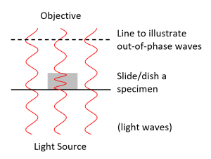
The refractive index of biological objects such as cells is generally higher than that of the surrounding medium (usually mostly water) meaning that light travels slightly slower through these objects. As light travels through the object (Fig. 1, gray rectangle), it slows down slightly until it exits the object, at which point it returns to its normal speed. Light that does not go through the object does not change speed. Although the light waves that do and do not travel through the specimen are ultimately going the same speed when they reach the objective, the light that goes through the specimen is a little bit behind (Fig. 1, dotted line). It’s like two identical runners that start a race at the same time, except that one runner has to run through a mud pit half-way through the race. The amount by which the light is slowed depends on the object’s refractive index and thickness. In the runner analogy, this is equivalent to saying that how much the runner is slowed depends on the thickness of the mud and the length of the pit.
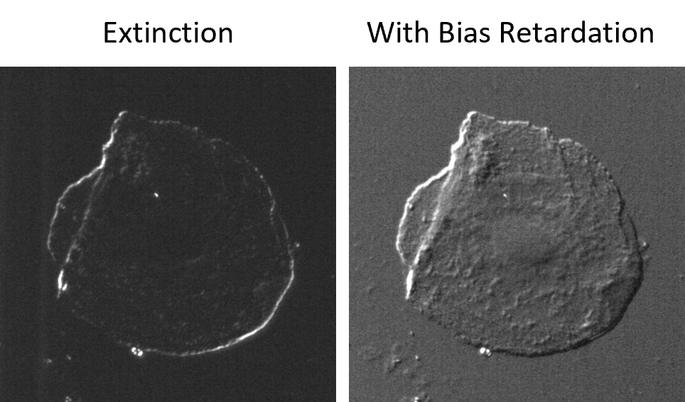
DIC helps us see the specimen by translating these light wave “delays” into differences in intensity. It tends to highlight the edges of objects (Fig. 2). You can change the contrast in your image by adjusting the bias retardation setting. At extinction, the edges of the objects are bright and everything else is dark. Adjusting the bias away from extinction gives the edges a shadowed appearance, making the object look 3D. Although the object may look like it is 3D, remember that the differences in intensity are due to the extent to which light waves are slowed, which is a function of an object’s thickness and refractive index. Therefore, the topography you see in your image is not simply a representation of thickness.
DIC does not work if there is any plastic in the light path (e.g. plastic coverslip, plastic lid on a glass-bottomed dish). DIC requires specialized microscope components: a polarizer and a prism with the condenser and a polarizer and prism with the objective. The prisms must be matched to the objective that you are using. In addition, the microscope must be aligned for Koehler Illumination. You will learn how to set-up DIC and adjust the bias retardation during your training.
